Go to the ONKODIN-Project (German)   German MDS-Forum 2010  |  Links  |  Contact us  |  Imprint  |  German Content German Content  
The Sysmex Scientific Calendar
Sysmex Scientific Calendar 2007
Lymphatic cells
Bacteria
Haemolysis
Acanthocytes
Inclusion bodies
Agglutination
Granulocytes
Malaria
Rare cells
Striations
Platelets
Cytoplasmic protrusions


Arte?-Fact? - Haemolysis - Calendar 2007 month 3

Microscopic differentiation always starts with low magnification screening of the blood film. This examination is used mainly to locate the appropriate area for microscopy, in addition to estimation of the cell count and inspection of the edge of the blood film. The erythrocytes should be lying next to each other in a monolayer, with only occasional contact and overlap.


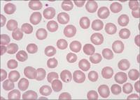
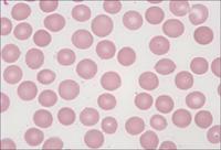
Erythrocytes of a normal person showing a paler centre and a darker border. Occasionally the pale centre has a elongated shape (stomatocytes, at this frequency they have no pathological significance).


The same blood film as above: At the edge of the blood film normal erythrocytes show no central pallor and look like sphaerocytes.


Pseudo-fragmentocytes (pseudo-schistocytes): artefacts due to mechanical abrasion close to a scratch.


Fragmentocytes (schistocytes) and thrombocytopenia in the case of a thrombotic thrombocytopenic purpura (TTP). In the partially thrombosed capillaries the erythrocytes are exposed to a high degree of shearing force which causes them to burst.


    Back to top of page Back to top                                                                                  Site Map Site Map  |  Printer friendly version of the document  |  Recommend this page!