Go to the ONKODIN-Project (German)   German MDS-Forum 2010  |  Links  |  Contact us  |  Imprint  |  German Content German Content  
The Sysmex Scientific Calendar
Sysmex Scientific Calendar 2007
Lymphatic cells
Bacteria
Haemolysis
Acanthocytes
Inclusion bodies
Agglutination
Granulocytes
Malaria
Rare cells
Striations
Platelets
Cytoplasmic protrusions


Arte?-Fact? - Inclusion bodies - Calendar 2007 month 5

During blood film inspection, and especially in the examination of erythrocytes, it is important to recognise the inclusion bodies and to distinguish between inclusions and artefacts.


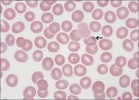
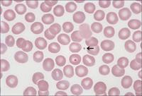
An apparent Howell-Jolly body, caused by contaminated microscope oil, can be identified as an artefact by the fact that it moves independently over time (see next picture).


The artefact in the microscope oil has moved further.


Large erythrocyte with basophile stippling (between the two lymphocytes) in a case of autoimmune haemolytic anaemia (AIHA). To the right, next to the lower lymphocytes, there is a polychromatic erythrocyte.


Reticulocyte staining. Characteristic HbH cell in a case of α-thalassaemia. Today, blood films are no longer investigated for HbH cells. Instead, α-thalassaemias are diagnosed by molecular genetic tests.


    Back to top of page Back to top                                                                                  Site Map Site Map  |  Printer friendly version of the document  |  Recommend this page!