Go to the ONKODIN-Project (German)   German MDS-Forum 2010  |  Links  |  Contact us  |  Imprint  |  German Content German Content  
The Sysmex Scientific Calendar
Sysmex Scientific Calendar 2007
Lymphatic cells
Bacteria
Haemolysis
Acanthocytes
Inclusion bodies
Agglutination
Granulocytes
Malaria
Rare cells
Striations
Platelets
Cytoplasmic protrusions


Arte?-Fact? - Striations - Calendar 2007 month 10

Staining must be standardised. Modification of the process, and also each new batch of staining solution should be controlled by a blood film made from normal blood: The colour of the erythrocytes must be salmon, the chromatin of the nuclei in the leukocytes must be clearly visible, and the cytoplasm of the normal lymphocytes must be blue. If with equal staining time the blood film or specific cells are different in colour, this has a diagnostic implication and is not detected as artefact.


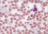
Striations with a blue cast and parts of the erythrocytes broken off, due to overheating of the blood sample (storage in the car with the sun shining on it).


A large number of flocculated cryoglobulins become visible when the condenser is lowered and no microscope oil is applied (compare with next picture).


When examined in the usual way with microscope oil the cryoglobulins appear as striations with a blue cast (the same sample and the same spot as above).


Reddish striations caused by extreme haemolysis in a case of septicaemia caused by Clostridium perfringens.


    Back to top of page Back to top                                                                                  Site Map Site Map  |  Printer friendly version of the document  |  Recommend this page!