Go to the ONKODIN-Project (German)   German MDS-Forum 2010  |  Links  |  Contact us  |  Imprint  |  German Content German Content  
The Sysmex Scientific Calendar
Sysmex Scientific Calendar 2008
Acute haemolysis
Schistocytes
Granulocytopenia
Extreme leukocytosis
Severe thrombocytopenia
Anaemia and erythrocytosis
Tumour cells
Acute leukaemia
Bacteria
Promyelocytic leukaemia
Thrombocytosis
Malaria
Reference scattergrams


The haematological emergency - Acute haemolysis - Calendar 2008 month 1

Haemolysis involves dissolution of erythrocytes by destruction of the cell membrane. At an elevated rate of haemolysis with an erythrocyte survival time of only a few weeks (standard is 120 days) the haemoglobin level may be kept stable or decline only slightly due to increased erythropoiesis (= compensated haemolysis). However, if the erythrocyte survival time is only a few days, even maximally elevated erythropoiesis cannot compensate for this loss, resulting in haemolytic anaemia. In case of acute intravascular haemolysis there is a risk of acute kidney failure due to free haemoglobin.


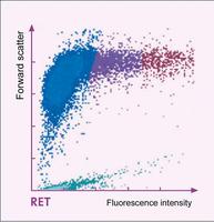
The increased synthesis of erythrocytes in the bone marrow results in an elevated number of reticulocytes (red and purple area) in the peripheral blood. It can be measured in the reticulocyte channel of the haematology analysers SYSMEX XE-5000, XE-2100 and XT-2000i. In this example the measured reticulocyte fraction was 7.9% (upper limit of the reference range: 2.1%). A better indicator of the increased erythropoiesis is the reticulocyte production index (RPI) which additionally considers haematocrit and reticulocyte maturation time.


Serum of the same patient as in the background figure at the time of hospitalisation (left) and 12 hours later (right): The serum is coloured red-brown due to intravascular haemolysis.


Autoimmune haemolytic anaemia (AIHA) in a case of chronic lymphocytic leukaemia (B-CLL): Reticulocytes are increased, spherocytes are present, Coombs’ test is positive.


Agglutinated erythrocytes and marked polychromasia in a patient with haemolytic anaemia caused by cryoglobulins in Waldenström’s macroglobulinaemia.


    Back to top of page Back to top                                                                                  Site Map Site Map  |  Printer friendly version of the document  |  Recommend this page!  
 
Internal Link
Reference scattergrams [More]